
Zygomatic Implants Before & After: What Results Look Like
After zygomatic implant treatment the change is functional first — a fixed upper bridge replacing a loose denture — and cosmetic second, with facial support often improving as a by-product. Before-and-after photographs are usually the first thing people look for and the hardest thing to interpret honestly. This page explains what actually changes, what the timeline from surgery to final teeth looks like, and how to read any clinic gallery critically before you let it influence a decision this significant.
- What changes first — and what does not
- How to read any clinic gallery critically
- The consent standards a clinic should meet
 Medically reviewed by Uzm. Dt. Oğuzhan SunarSpecialist PeriodontistUpdated August 2026 · 9 min readWritten from 4 cited sources · clinical sign-off pendingPublished by Natural Clinic · how this guide is funded
Functional change
Comes first
Illustrative photography — not a patient of any clinic.
Functional change
Comes first
Final bridge
4–6 months
indicative
Patient photos
Consent required
Results
Vary by individual
What "before and after" really means here
Most people arriving at this page are not comparing smiles out of curiosity. They have been told there is not enough bone for conventional implants, they have lived with a denture that moves, and they want to know what the other side of treatment looks like. That deserves a better answer than a grid of photographs.
The change that matters most is rarely the one a camera captures. Zygomatic implants replace a removable appliance resting on shrinking gum with a bridge fixed to the cheekbones. The visible difference is a set of teeth; the difference patients describe is biting into food, speaking without a denture shifting, and stopping thinking about their mouth altogether. Function first, appearance second — in that order, and on different timescales.
A photograph also cannot show whether the bite was comfortable, whether speech settled, whether the case was straightforward or a difficult revision, or whether anything went wrong. For that side of the picture, read our page on success rates, risks and complications.
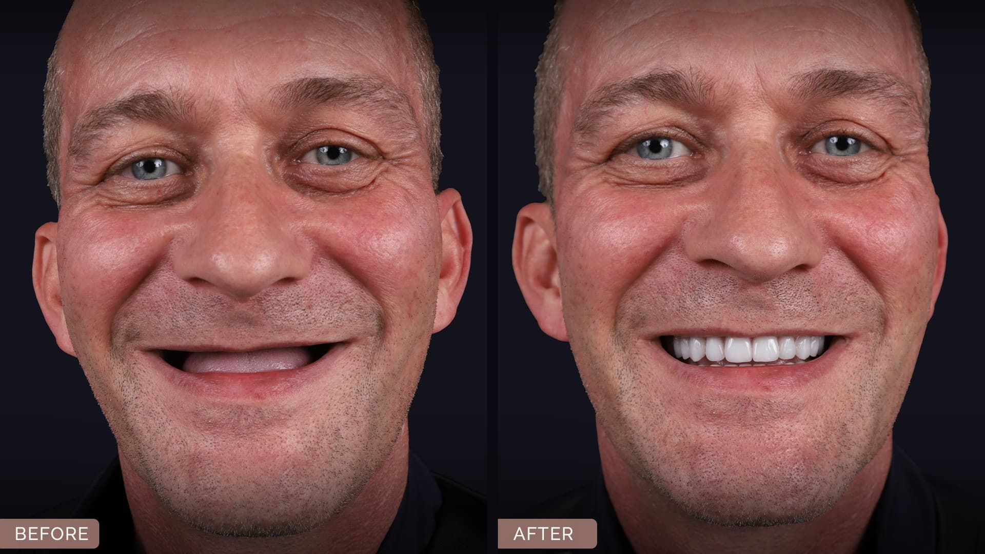 Real patient case · consent confirmed
What changes first: function
A fixed provisional bridge is normally attached within days of surgery, and the first differences patients describe are practical rather than cosmetic: nothing to take out at night, nothing lifting when you laugh, no adhesive in your morning routine.
Chewing changes more gradually. A soft-food period applies while the implants integrate, so biting into firm food generally arrives with the final bridge rather than immediately after surgery — anyone promising a date for that is guessing. Our recovery guide sets out the stages. Speech usually runs the other way: a new bridge changes the shape of the palate, so altered "s" sounds or extra saliva are common at first and settle over the following weeks. If they do not, the prosthetic design can often be adjusted, so raise it.
Before and after, described rather than photographed
The changes patients and clinicians most often describe. These are general patterns in denture-to-implant treatment, not a prediction for your case.
Before: a failing upper denture
Best for: Where most candidates start
- Eating
- Firm and chewy foods avoided; chewing efficiency reduced
- Stability
- Lifts or rocks; adhesive relied on; relines needed as bone shrinks
- Speech
- Clicking or slipping during conversation is common
- Comfort
- Sore spots and pressure areas on the gum
- Confidence
- Many patients report avoiding eating out or public speaking
- Bone
- Upper jaw continues to resorb under the denture over time
After: a fixed full-arch bridge
Best for: What treatment aims to achieve
- Eating
- A wider range of foods for most patients once the final bridge is fitted
- Stability
- Screw-retained onto the implants and not removed by the patient
- Speech
- An adaptation period is normal; design can be adjusted if it does not settle
- Comfort
- No gum-borne pressure, though implant-site tenderness can occur early
- Confidence
- The change most often described by patients, and the hardest to photograph
- Maintenance
- Daily cleaning under the bridge plus scheduled reviews, for life
What changes second: appearance and lip support
Facial appearance does change for some patients, but the honest version is more modest than marketing suggests. Long-term tooth loss and bone resorption can shorten the lower face and reduce support behind the upper lip, and a well-designed full-arch bridge can restore some of that. Where it happens, patients tend to describe looking rested rather than looking different.
Two limits are worth stating plainly. The lip support achievable is dictated by prosthetic design and your own anatomy rather than by the use of implants — a well-made denture can also provide lip support, and a poorly designed fixed bridge can provide less. And although zygomatic implants are anchored in the cheekbone, they do not build up or reshape it: this is not a facial aesthetic procedure, and any suggestion that it lifts the cheeks should be treated with scepticism.
Tooth shade and shape, how much gum shows when you smile and how full the lip looks are prosthetic decisions. They should be discussed and, where possible, previewed before the final bridge is made rather than presented as a surprise at fitting.
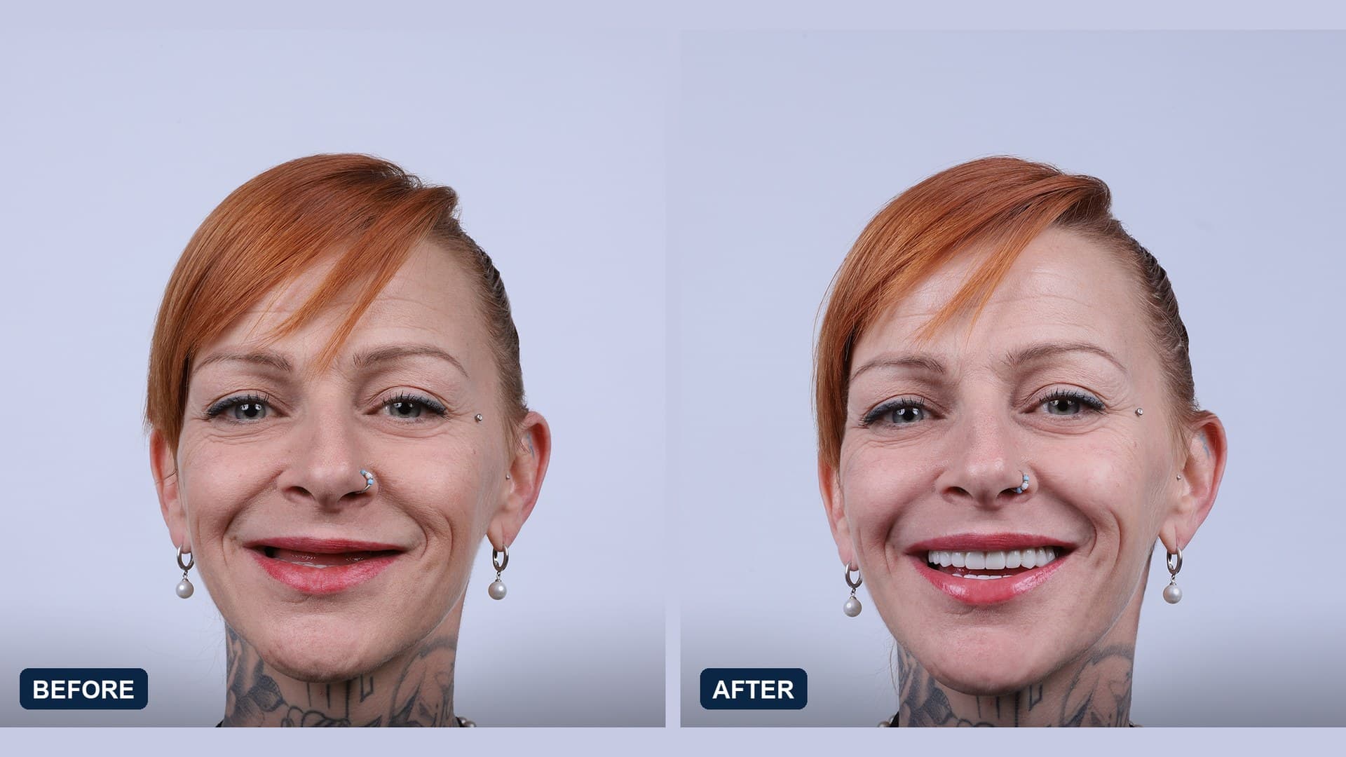 Real patient case · consent confirmed
From surgery to final teeth: when the "after" is taken
This sequence is the key to reading any gallery, because the same patient looks different at every stage. Protocol details vary between clinics and cases.
- 1
Before: the documented starting point
Full-face and smile views, intra-oral photographs and the CBCT scan. These are a clinical baseline, not marketing material — and a gallery without a genuine "before" has no comparison to offer.
- 2
Surgery and the fixed provisional bridge
A provisional bridge is normally attached within days. Photographs here show a temporary prosthesis on a swollen, bruised face, which is why most clinics do not publish them.
- 3
The swelling phase
Swelling and bruising over the cheeks, and sometimes around the eyes, are expected after this surgery and settle over the following weeks; timescales differ and no honest clinic will promise a date. Nobody looks like their final result here.
- 4
Integration, wearing the provisional bridge
A months-long period of integration on a soft-food diet. Many published "after" photographs are actually taken during this phase with an acrylic provisional in place — a legitimate stage to photograph, but one that should be labelled.
- 5
The final bridge
The definitive bridge is designed, tried in and fitted. This is the only fair "after" for judging shade, tooth position and lip support, and the point at which a full diet normally returns.
- 6
One year and beyond
The most informative image and the rarest: the same patient a year or more on, soft tissue settled and the bridge in real use. A clinic that publishes long-term follow-up is telling you something meaningful.
Why photographs vary so much between clinics
Two galleries can show identical clinical work and look nothing alike, because much of what you respond to is photography rather than dentistry. Lighting direction, flash, lens, head angle, lip retraction, a posed versus natural smile, editing and even a change of lipstick all shift the result.
The pattern is easy to spot once you know it: the "before" taken under harsh overhead light, unsmiling, with an ill-fitting denture; the "after" taken with soft directional lighting, a rehearsed smile and sometimes visible retouching. That is a photographic transformation layered on a clinical one, and it makes cases look more dramatic than the treatment can claim credit for.
This does not make galleries worthless. It makes them evidence about a clinic's documentation standards as much as its surgery — and a clinic that photographs consistently, labels the treatment stage and shows unflattering angles is telling you something reassuring about how it works.
How to read a before-and-after gallery critically
Use this on any clinic's cases — including cases published here once consented imagery is available.
Last updated: July 2026| What you see | What it may mean | What to ask |
|---|---|---|
| Different lighting or background between the two images | Part of the apparent improvement is photographic, not clinical | Do you have matched-lighting photographs of this case? |
| Head tilted or angled differently | Angle alters apparent tooth position, lip fullness and face shape | Can I see standardised full-face and profile views? |
| No treatment stage stated | The "after" may show a temporary bridge with swelling still settling | Is this the provisional or the final bridge, and how long after surgery? |
| Unnamed, undated, unsourced cases | The case may not be the clinic's own work; stock and reused images circulate widely in this sector | Was this treated by the surgeon who would treat me, in this clinic? |
| Only wide smiles, never a relaxed mouth | A relaxed lip reveals tooth position and the bridge transition; a broad smile can conceal both | Can I see this patient at rest as well as smiling? |
| Every case looks flawless, and teeth are uniformly white | Publication is selective everywhere, and retouching makes clinical assessment impossible | Are these unedited apart from cropping, and can you show me a case that did not go to plan? |
Photographs are not evidence of outcome quality and are never a substitute for a CBCT-based clinical assessment of your own case.
Questions to ask about any case you are shown
If a gallery is influencing your decision, these questions establish whether it should.
- 1Was this case treated by the surgeon who would treat me, in the clinic I would attend?
- 2Which configuration was used — hybrid zygomatic, or quad zygoma?
- 3Was the upper arch treated alone, or both arches?
- 4Is the "after" image the provisional bridge or the final bridge, and how long after surgery?
- 5Were both photographs taken with the same lighting, camera and head position?
- 6Has the patient given written consent for these images to be published?
- 7Do you have follow-up images of this case at a year or more?
- 8Did this patient have any complications? See risks and complications.
What results tend to look like in different situations
Starting points differ enormously, which is the main reason results do. These are general patterns, not predictions.
Long-term denture wearer moving to fixed teeth
Quad zygoma results — almost no usable upper jawbone
After failed implants or a failed bone graft
Zygomatic combined with pterygoid implants
Upper arch only, or both arches
The consent and honesty standards a reputable clinic meets
These are the standards we hold our own future gallery to, and a reasonable benchmark for anyone else's. UK advertising and professional guidance on patient imagery applies to any clinic marketing to UK patients, wherever it is based.
- Written, specific, documented consent for publication — not consent buried in a treatment agreement
- Consent that can be withdrawn, with images removed when it is
- Its own cases only, never purchased or stock imagery presented as clinical work
- The treatment stage stated: provisional bridge or final bridge
- The interval since surgery stated for each image
- The configuration stated — hybrid, quad zygoma, and which arches were treated
- No retouching beyond cropping, and no whitening or smoothing of teeth or gums
- Consistent lighting, camera and head position between before and after
- A visible "individual results vary" statement beside the images, not hidden in a footer
- Unsuccessful or complicated cases discussed openly at consultation, even if not published
- Compliance with UK-GDPR when patient images and records are handled or transferred
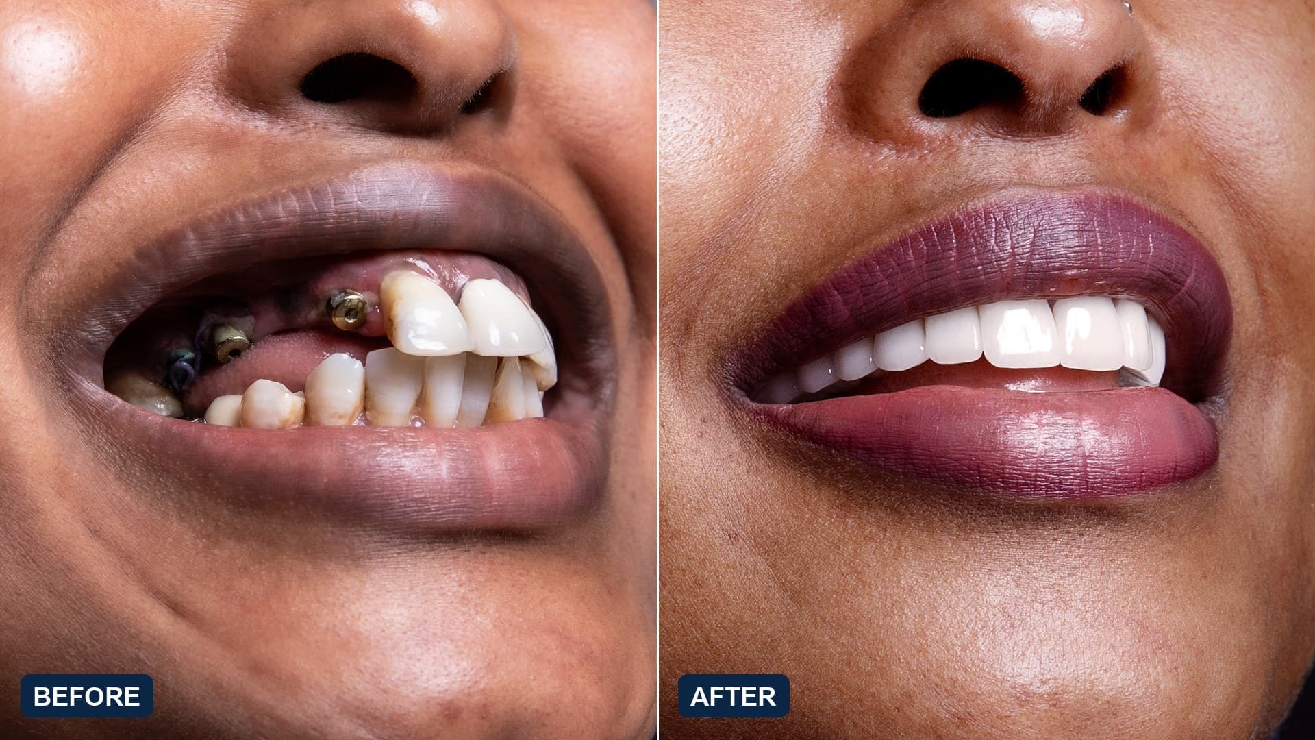 Real patient case · consent confirmed
Frequently asked questions
Why are there no before-and-after photographs on this page yet?
What changes first after zygomatic implant treatment?
Will zygomatic implants change my face?
How long until I see the final result?
How can I tell whether a clinic's before-and-after gallery is trustworthy?
What is the difference between quad zygoma results and standard zygomatic results?
Can I see results specifically for moving from dentures to implants?
How we reviewed this page
Written by the ZygomaticImplant.net editorial team and checked against the published literature. What we accept as a source, the difference between a page that is cited and one that is clinically signed off, and how we handle corrections are all set out in our editorial standards.
- First published
- 10 August 2026
- Last revised
- 21 August 2026
- Cited sources
- 4
- Clinical sign-off
- Pending · 25 statements queued
Sources & references
- [1]Advertising Standards Authority / CAP Code guidance on advertising dental treatment, including the rules that apply to before-and-after imagery and health claims.
- [2]Davó R, Fan S, Wang F, Wu Y. Long-term survival and complications of Quad Zygoma Protocol with Anatomy-Guided Approach in severely atrophic maxilla: A retrospective follow-up analysis of up to 17 years. Clin Implant Dent Relat Res. 2024;26(2):343–355. Context for what a long-term result actually looks like: 97.8% implant survival over a mean 8.8 years, but with local orofacial inflammation in 35.7% of patients and sinusitis in 12.5%, mostly arising years after surgery.
- [3]Linn TT, Khaohoen A, Thu KM, Rungsiyakull P. Oral-Health-Related Quality of Life in Elderly Edentulous Patients with Full-Arch Rehabilitation Treatments: A Systematic Review. J Clin Med. 2024;13(12):3391. Implant-supported prostheses produced clearly greater improvement in oral-health-related quality of life than conventional dentures.
- [4]Al-Nawas B, Aghaloo T, Aparicio C, et al. ITI consensus report on zygomatic implants: indications, evaluation of surgical techniques and long-term treatment outcomes. Int J Implant Dent. 2023;9:28. Mean survival 96.2% over a mean 6.3-year follow-up; sinusitis the most common complication at 14.2% prevalence. Individual results shown on this page are single cases and are not a prediction for anyone else.
- [5]General Dental Council guidance on advertising, patient consent and the use of patient images — exact current reference still to be added.pending confirmation
- [6]UK-GDPR requirements for processing patient images and clinical records, including cross-border transfer — current reference still to be added, and this page should not be relied on for the legal position.pending confirmation
- [7]Case details, treatment dates and the interval between photographs for the consented cases shown here — still to be confirmed in writing by the treating clinic. The ASA before-and-after rules require an accurate statement of the time elapsed, so no interval is claimed until that is supplied.pending confirmation
- [8]This page is for information only and is not a substitute for professional medical advice, diagnosis or treatment. Some clinical statements are still awaiting sign-off by our medical reviewer.
The next step is a scan, not a decision
Upload a panoramic X-ray or CT scan and a specialist will tell you what is actually possible in your case — in writing, with no obligation.
Medically reviewed · Free · No pressure, no spam
Related guides

Zygomatic Dental Implants: The Complete Guide
Read
The Zygomatic Implant Procedure, Step by Step
Read
Zygomatic Implant Recovery & Aftercare
Read
Zygomatic Implants: Success Rates, Risks & Complications
Read
Quad Zygomatic Implants: Fixed Teeth With No Usable Jawbone
Read
Zygomatic Implant Costs: UK vs Abroad
Read
Dentures vs Zygomatic Implants: Making the Switch
Read